|
Machine Vision News
Vol. 4, 1999
|
"Crystal clear!"
Real-time image processing and visualisation brings medicine
into the new age
With the increasing demand for minimally
invasive surgery, MRI scanners have a major part to play not only as a diagnostic
tool but also as means of guiding the surgeon´s hand. However, before MRI scanners
can be used in an interventional role, certain technological problems must be overcome.
A multidisciplinary consortium that brings together Oulu Hospital, the Electrical
Engineering Faculty at Oulu University, Daum GmbH, an innovative German engineering
company, and Picker Nordstar Oy, a global leader in the medical imaging industry,
have been working together on the IRVIT project to solve exactly those problems.
Background
Radiology is the field of medicine that
uses various forms of acoustic and electromagnetic energy for producing diagnostic
images of the human body. Since the discovery of X-rays by Rontgen in 1895, X-ray
imaging has dominated the history of radiology and it has only been during the last
three decades that alternative imaging methods have begun to emerge. The first of
these was ultrasound, which used acoustic waves in forming an image. Shortly afterwards
computed tomography revolutionised the world of medicine when it made it possible
to acquire cross-section images from a living human body. One drawback, however,
was that computed tomography still used X-rays, which are harmful to living organs.
In the late 1970s magnetic resonance imaging
(MRI) was introduced. MRI is able to produce two- or three-dimensional images of
great quality, showing cross-sections through body parts at regular intervals. The
images are so precise that radiologists are often able to get as much information
from a scan as from looking at the tissue directly. The MRI scanner uses a modulated
magnetic field and radio frequency (RF) energy to ´excite´ the hydrogen protons
of organic tissue. When the protons ´relax´ again, they send out an RF signal which
is measurable. Using special measurement techniques, the proton relaxation signal
can be interpreted into a cross-section image of a human body.
MRI is the state-of-the-art radiological
imaging method that has far superior soft tissue contrast to any other radiological
imaging methods. What is more, in contrast to X-ray based methods, it has the major
advantage of providing images without the use of harmful ionising radiation. Not
surprisingly, its applications have grown rapidly in recent years.
Interventional radiology
Interventional radiology uses imaging to
guide surgical operations. It is part of the recent trend of so-called keyhole surgery,
which aims at minimal invasiveness. Obviously when operations are performed through
such small openings, image guidance is needed. For example, by using ultrasound
or X-ray fluoroscopy, a needle can be guided to the chosen part of a tissue and
inject medication into or take a tissue sample from a tumour. As medical technology
has improved, the distinction between interventional radiology and surgery has become
fuzzier.
Currently 99.5% of MRI scanners are sold
for diagnostic imaging. However the recent development of the new open magnet systems
has made ´access´ to the patient during imaging possible. This development means
that there is now the potential to combine state-of-the-art imaging with surgical
or radiological interventions.
Performing surgical operations however,
is not straightforward: the room or environment where the MRI scanner is located
has extremely strict instrumentation demands. For example, any devices and instruments
brought into the MRI room must not interfere with the image acquisition of the scanner.
In other words, they should neither emit RF energy nor should they distort the magnetic
field inside the imaging volume. At the same time, though, the devices and instruments
should themselves be able to withstand strong magnetic field and RF emission.
If interventional MRI is to become a surgical
reality, a great deal of development work is required. New instruments need to be
designed, operating room instrumentation will have to be altered to meet MRI requirements,
and the scanners themselves will need to be modified to raise the image acquisition
to a more interactive level. Many scientific fields and techniques will be required
in the realisation of this goal, and naturally modern image processing technology
will have a significant role to play.
IRVIT demonstration
Intraoperative Real-time Visualisation
and Instrument Tracking (IRVIT) is an EU-funded research project under the HPCN-TTN
initiative.
The IRVIT project target is to demonstrate
how innovative technology can be used in interventional MRI. The two key features
to be demonstrated are:
- accurate instrument tracking
- real-time image processing and
visualisation.
The problems posed by interventional MRI
are very similar to those of the better known field of telemanipulation. The MRI
intervention system is a closed feedback loop where the surgeon moves an instrument
according to the visual feedback he gets. Moving the instrument triggers an image
update, which shows the instrumentÕs new position with respect to the anatomy. In
order to be able to control the image acquisition and thereby provide visual feedback,
it is necessary to be able to assess the location of the instrument accurately.
This is where the IRVIT project comes in.
A new technological device, the ESR marker, has been developed and this can be used
to measure the location of the surgical instrument. The marker is a very small crystal
that can be integrated into the tip of an instrument 1.2mm in diameter. By measuring
the electron spin resonance (ESR) signal from the crystal, the location of the instrument
can be etermined. As the MRI scanner is used to measure both the ESR marker location
and the natomical images, most of the sources of error will be the same, and thus
their net effect is cancelled out. As a result excellent relative localisation accuracy
can be achieved.
Figure 1:
ESR technology provides an accurate
and reliable method of instrument location,
even when the instrument bends.
|
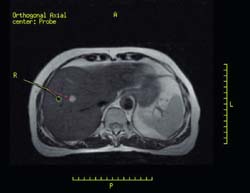
|
During an intervention the images are almost
always acquired relative to the tip of the instrument. There exist methods that
can measure the location of the non-invasive part of the instrument body. With these
systems the tip of the instrument is extrapolated and thus bending of the instrument
can dramatically impair accuracy. Because the ESR marker is small, it can be located
directly into the tip of the instrument. Therefore instrument bending does not impair
accuracy. An example of a case where ESR marker tracking is used is shown in Figure
1.
The images obtained by the scanner itself
are insufficient for the efficient performance of a surgical intervention and various
image processing techniques are necessary in order to make the most important features
more easily visible. In addition, real-time volume rendering techniques can provide
essential added value to the visual feedback. The applications of these technologies
are currently being studied at Oulu University Hospital.
Another important image processing task
in IRVIT is the registration of real-time images with earlier preoperative images.
The current image acquisition latency of the MRI scanners is a result of the limits
of imaging physics and of the MRI scanner design. In diagnostic use, the image acquisition
latencies do not play as critical a role as they do in interactive interventional
MRI. MRI scanner manufacturers are currently working on eliminating both sources
of latency but for the present and near future, real-time imaging is not yet a possibility.
Fortunately this problem can be alleviated if the preoperative images can be used
parallel to the intraoperative ones. The research into image registration methods
now being done at the University of Oulu aims to solve this problem. A demonstration
system of the IRVIT project will be built into the Picker Outlook Proview scanner
at Oulu University Hospital in Finland. This demonstrator will be available for
evaluation at the end of June 1999, when the project is due for completion. Figure
2 shows a preview of the forthcoming system.
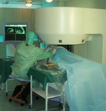 Figure 2: Caption: A radiologist
testing the IRVIT demonstrator system.The Consortium
Figure 2: Caption: A radiologist
testing the IRVIT demonstrator system.The Consortium
Any project in the field of interventional
MRI requires a multidisciplinary consortium and skills, and thus the consortium
of the IRVIT project consists of four complementary partners, each leaders in their
respective fields.
Daum GmbH is an innovative German engineering
company which specialises in developing instruments for minimally invasive surgery
and interventional radiology. In a very short time, its high quality MRI-compatible
instruments have become well known all over the world. Within the IRVIT project,
Daum has used its substantial expertise in MRI-compatible materials and instruments
to integrate the ESR marker into an instrument.
Picker Nordstar Oy is a subsidiary of Picker
International, a global leader in the medical imaging industry. Its products include
Outlook Proview, one of the MRI systems with the most open shape available on the
market today. With a large patient gap (i.e. there is sufficient space for the surgeon
to work around the patient) and flexible patient handling, Outlook Proview offers
the ideal platform for developing interventional procedures. The Outlook Proview
scanners are designed and manufactured in Finland.
The Department of Radiology at Oulu University
Hospital is one the leading clinics involved in MRI research and interventional
radiology in Northern Europe. Not only does the clinic have over ten engineers and
physicists researching MRI imaging, image processing and real-time volume endering,
but the modern interventional MRI facilities at the hospital are ideal for performing
and developing both radiological and neurosurgical procedures. The Department of
Radiology therefore brings significant accumulated expertise in state-of-the-art
image-guided procedures to the IRVIT project.
The Department of Electrical Engineering
in the University of Oulu has a Machine Vision and Media Processing Group with over
60 engineers and mathematicians specialising in signal, image and media processing.
This group has applied its expertise in image processing to the specific applications
of the IRVIT project.
Contacts
Technical co-ordinator of IRVIT
Lasse Jyrkinen
Picker Nordstar Oy
Tel: +358-8-553 2787
Fax: +358-8-553 2612
E-mail: lasse.jyrkinen@oulu.fi
TTNMV-SF (URL)
http://www.vtt.fi/ttn
HPCN-TTN network (URL)
http://www.hpcn-ttn.org/
|