Education: Digital X-ray

Over the last few years, as manufacturers and researchers
around the world have taken digital x-ray detectors out of the lab and into clinical
trials, it's become clear that digital x-ray imaging will indeed revolutionize diagnostic
radiology.
In fact, many observers have hailed it as the most significant breakthrough in x-ray
imaging in the last 25 years, referring to it as "the New Modality."
No matter how large or small your facility may be,
it's time to start giving serious consideration to how digital detection will impact
your department and which technology will meet your needs most thoroughly and cost-efficiently.
This tutorial introduces the key concepts around Digital
X-ray technology and analyzes its impact and potential benefits.
Part 1: Overview
From four Components to one
The promise of Digital X-ray
Improving diagnostic confidence
Bottom-line benefits
Part 2: the different Digital X-ray technologies
The GE approach
Alternative approaches to digital x-ray
Part 3: Current Applications
Full Field Digital Mammography
Digital Radiography
All-Digital Cardiology
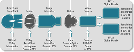
To understand the potential significance of digital
detectors, consider what happens to the x-rays collected by even a state-of-the-art
conventional x-ray device, such as a radiography & fluoroscopy system equipped
with a digital box: The x-ray signal is transmitted from the tube, through the patient,
and into an image intensifier. Next, the signal is processed through image-intensifier
and camera optics before being sent on to a video camera and through digitization
(Fig. 1a). Finally, the remaining signal is sent on for display and hard-copy generation.
Note that at each stage in this process, the x-ray
signal is degraded to some extent, even if the individual components are optimized
for the application. As a result, typically less than 40% of the original image
information is available for use in image production.
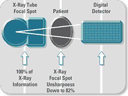
Now consider what happens when we add a digital detector
to the equation: It replaces everything but the x-ray tube and patient (Fig. 1b)!
Because of its high Detective Quantum Efficiency (DQE), it has the potential to
capture over 80% of the original image information. And it equips the user with
a wide range of post-processing tools to further improve that signal ' including
many that can be applied automatically.
 Fig. 1a:
In a conventional, digitized R&F imaging chain, the signal degradation that
occurs with each component consumes more than 60% of the original x-ray signal.
 Fig. 1b:
A digital detector replaces all these components, allowing the user to preserve
more than 80% of the original signal ' and to further enhance that signal automatically
or explicitly.
What's more, these advantages are applicable to the
full range of x-ray imaging modalities, including chest and breast imaging, and
in the future fluoro-based applications.
The result? Far more accurate and detailed renderings
of the anatomy of interest ' and a wealth of opportunity for further enhancing the
diagnostic utility of each and every exam you conduct.
The advent of digital x-ray technology means that
the world's Radiology departments will soon participate in a transformation of revolutionary
proportions, and realize revolutionary advantages. Imagine the benefits of routinely:
- Processing image data to highlight regions of interest
and suppress irrelevant information, thereby improving the diagnostic utility of
virtually any study
- Combining image data with other pertinent patient information
available from RIS/HIS systems
- Quickly transmitting the resulting files anywhere over
the networking connections of your choice, including conventional phone lines
- Archiving all this information in minimal space, and
retrieving perfect originals at the touch of a few keys
As already reported by some sites, it won't be much
longer before capabilities such as these become available to all Radiology departments
around the world.

|
Click on image to enlarge
Fig. 2: Magnified Contrast Detail Phantom Images
Left: Images acquired on Film
Right: Images acquired with a Digital detector allow detection of lower contrast
objects than conventional film images
|
Some of digital-detector technology's most obvious potential
advantages fall into the clinical realm.
For example, digital imaging is already proving its
ability to enhance both radiographic and fluoroscopic image quality by improving
latitude, DQE and contrast resolution (Fig. 2).
It's permitting further improvements in the diagnostic
utility of virtually any study through advanced image post-processing techniques.
And it has the potential for lowering patient radiation
dose, not only during the initial exam, but also by minimizing the need for retakes.
The result: improved diagnostic confidence for a full
range of applications.

|
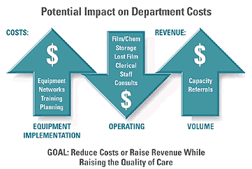
Fig. 3: Potential Impact on Department costs
Over time, departments using Digital-detector technology stand to realize substantial
financial rewards.
|
We can now predict with certainty the financial ramifications
of the Digital X-ray technology. It already seems clear that users will reap substantial
bottom-line rewards as digital technology takes hold, for reasons such as these
(Fig. 3):
- Operating costs should decrease because of dwindling
requirements for film, processing equipment, chemicals, and archiving space ' not
to mention the labor now required for managing these activities.
- Thanks to the inherent speed of digital exams, increased
patient throughput should drive up revenues for facilities with adequate patient
volume ' or, alternatively, allow reductions in the number of rooms and their associated
costs.
- Properly engineered digital detectors may pave the way
for a broad range of future advanced applications, such as Computer Aided Detection
and teleradiology, to permit cost-cutting consolidation of resources.
|