- What is color?
- How do we see in color?
- What are color vision
deficiencies? How common are they?
- What does it mean to
be color normal
- What parts of the eye
are important for color vision?
- What color processing
takes place in the eye? the brain?
- What spatial and temporal
processing takes place in the eye? the brain?
- What is chromatic adaptation?
- How do we characterize
a person's color vision?
- How do we select names
for colors?
- How many colors can
we see?
- What is object color?
What other modes are there?
1. What is color?
Color is that characteristic of a visible object or light source by which an observer
may distinguish differences between two structure-free fields of the same size and
shape, such as may be caused by differences in the spectral composition of the light
concerned in the observation.[ref
1, p 723] In other words, color is that perception by which we can tell
two objects apart, when they have otherwise similar attributes of shape, size, texture,
etc.
OK, that's the textbook answer. This is admittedly unsatisfying, because color is
an inherently subjective experience. Color only exists in our minds, and putting
a scientific definition together of no easy task. The usual definition, given above,
is really a circular argument. It amounts to: "Color is that attribute of an object
leftover when you eliminate all attributes except color." So, if an two objects
look different, but have the same size, shape, texture, etc., then the way you are
telling them apart is their color. Still not satisfied? Here's
another answer for you.
Back to top
2. How do we see in color?
In the retina of our eye are photoreceptors that are sensitive to light. When light
is absorbed by the photoreceptors, the light energy is converted into electrical
and chemical signals that the neurons in our eye and brain process. There are two
kinds of photoreceptors in the retina: rods and cones. Rods mediate vision at lower
levels of illumination. Cones mediate vision at higher levels of illumination. There
are three types of cones with each type differentially sensitive to a different
region of the visible spectrum. They are known as the Short-wavelength sensitive
cones, the Middle-wavelength sensitive cones and the Long-wavelength sensitive cones.
Sometimes they are referred to as R-, G-, and B-cones but these are misnomers based
on the colors in the spectrum. For example, very short wavelength light can uniquely
stimulate the S-cones but the sensation associated with this light stimulation has
a reddish and bluish component. Fundamentally our color vision derives from comparisons
between the amount of light being absorbed by each cone type. Our visual system
compares the outputs of the cone types to process color. In addition, color appearance
is influenced by the ratios of cone excitations in surrounding regions and by the
overall levels of cone excitation caused by the prevailing illumination. These comparisons
occur at different stages of processing that start in the retina and continue to
the cerebral cortex of the brain.
Back to top
3. What are color vision deficiencies? How common are they?
Color vision deficiencies result from either a lack of one or more cone types, or
cones that behave somewhat differently from average. Those lacking long wavelength
cone pigment suffer from protanopia. A related condition is anomalous protanopia,
or protanomaly. Here, long wavelength cones are present, but their sensitivity is
shifted spectrally to shorter wavelengths, so they interpret certain stimuli differently
than normal observers. Similarly, deuteranopia (deuteranomaly) is the lack (spectral
shift to longer wavelengths) of the middle wavelength cone pigment, and tritanopia
is the lack of short wavelength cones (tritanomaly is incomplete tritanopis). It
is important to remember than none of these conditions should be referred to as
true color blindness. This is not simply a politically correct statement. In fact,
those suffering from any of these conditions do experience color, but not in the
sense that a color "normal" observer does.
Other less-common deficiencies are rod monochromacy and cone monochromacy. With
rod monochromacy, there are no cones present, only rods. Persons suffering from
this are truly color blind. With cone monochromacy, a person has only one cone type.
For more details on these and other vision deficiencies, see
reference 3.
From reference 2,
percentages of the more common deficiencies:
|
Type
|
Male %
|
Female %
|
|
Protanopia
|
1.0
|
0.02
|
|
Deuteranopia
|
1.1
|
0.01
|
|
Tritanopia
|
0.002
|
0.001
|
|
Cone monochromatism
|
~0
|
~0
|
|
Rod monochromatism
|
0.003
|
0.002
|
|
Protanomaly
|
1.0
|
0.02
|
|
Deuteranomaly
|
4.9
|
0.38
|
|
Protanomaly
|
~0
|
~0
|
|
Totals
|
8.0
|
0.4
|
4. What does it mean to be color normal?
A color normal individual is one whose color vision is not greatly different from
the average person. This may seem obvious, but remember that the color vision deficiencies
are, in most cases, not simply on/off conditions. There is a continuum of, for example,
deuteranomalous people. Some will have only a slight shift in middle-wavelength
cone sensitivity and others may have so large a shift that middle-wavelength cone
behavior is no longer distinguishable from that of long-wavelength cones. A series
of color-matching experiments can determine what type of deficiency a person might
have. The end result is usually simply an understanding of approximately how far
from average an individual might be. To state a cutoff between normal and anomalous
vision assumes some criterion which is application specific. In other words, just
how normal you are depends on just how normal you need to be for your task. If you
are a taxi driver, you certainly need to determine the color a traffic signal. If
you are an interior designer, you probably need somewhat better color vision to
please your customers.
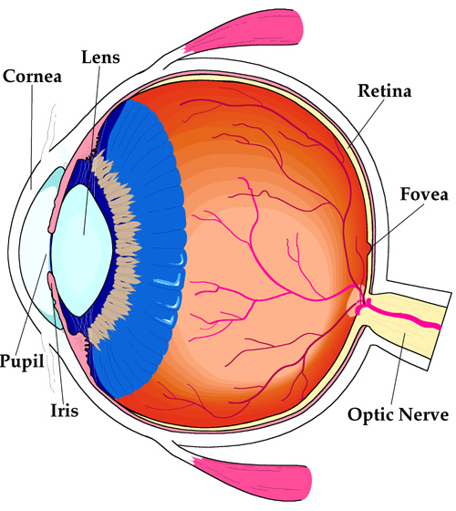
5. What parts of the eye are important for color vision?
 Click on the image for a larger view
Use by permission from
reference 4.
|
Most of the important parts of the eye are labeled on the diagram. The cornea and
lens focus the image onto the retina. The retina is the part that actually detects
incoming light. The iris adjusts in width to partially account for light levels.
The fovea is the central focal point of the eye. (That is, when we look at something,
we are casting its image onto the fovea.) The fovea is the are where we get most
of the spatial detail and color in what we see. The optic nerve is a bundle of nerves
which carries the visual information to the brain. Not shown is the macula, which
is a filter over the fovea. It serves to limit the damage that might be cause to
the fovea if we accidentally focus on intense light sources, such as the sun.
|
Back to top
6. What color processing takes place in the eye? the brain?
During the development of the embryo, part of the neural tube which develops into
the central nervous system forms outcroppings that extend and develop into the retinas.
Therefore the retina is considered part of the brain: a part that is easily accessible
for study. The retina consists of 5 layers of cells and between each layer there
are extensive interconnections in which visual processing takes place. There are
about 125 times as many receptors in the retina as there are ganglion cells in the
optic nerve, which connects the eye to the brain. This gives an indication of some
of the processing that must go on. Because the retina is so accessible, much is
know about the processing of color. The signals from different cone types are segregated
in opponent pairs during this processing so that ganglion cells have specific receptive
fields that are excited by one cone type in the center and inhibited by another
in the surround. Although the effects this organization can be evidenced in psychophysical
experiments that measure different aspects of visual function, our conscious experience
of color does not relate well to this organization. It is at the later stages of
processing in the brain in which our subjective experience of color is processed.
Back to top
7. What spatial and temporal processing takes place in the eye? the
brain?
Subjectively, we experience many different qualitative aspects of vision, for example,
form, color, motion, depth, etc. This information is all input into the visual system
through the photoreceptors in the retina. Therefore the retina has to process the
information to preserve all these different aspects of the visual stimulus. The
term multiplexing is often used to describe the way the early visual system simultaneously
sends this varied information to the brain. Specialized pathways are determined
very early in visual processing. The second layer of retinal cells already has specialized
neurons that process form by creating center-surround receptive fields. Different
types of temporal response are seen in cells with either sustained or transient
responses in the next layer of processing. Two streams of visual processing have
been identified at the optic nerve level. One responsible for fine detail and color
and the other responsible for detecting rapid temporal change with high sensitivity
to changes in contrast. (In other animals, cells that respond to directional motion
are already present in the retina.)
In the brain we see evidence of hierarchical and modular processing of visual information.
At the early stages of cortical processing, cells exist that respond to stationary
or moving edges and bars of light contrast, evidence of form processing. At later
stages of cortical processing we find areas of the brain that seem to be specialized
for the processing of specific visual attributes. For example area MT in the temporal
lobe shows a specialization for motion detection. Not only do cells here respond
best to moving stimuli with specific characteristics (direction and speed) but also
studies with animals has demonstrated that artificially stimulating these cells
can influence the perceptual judgments of motion. In monkeys, an area called V4
has been identified as being critical for the processing of color. It has been shown
that humans with brain damage in the area homologous to this one lose color vision.
Back to top
8. What is chromatic adaptation?
Chromatic adaptation is the ability of the human visual system to adjust itself
in response to varying illuminant conditions. In other words, we adapt to the color
of the light source in order to better preserve the color of objects. For example,
if viewed under incandescent light, white paper has a decidedly yellow cast. However,
we have the ability to automatically account for the yellowish light, and we therefore
see the paper as white. If you think about it, this makes a lot of sense. It would
be a very confusing world if objects were changing color every time the light source
changed. From an evolutionary point of view, we still need to know if the fruit
is ripe whether it is morning, noon, or evening. Chromatic adaptation makes this
possible.
Back to top
9. How do we characterize a person's color vision?
There are several test available, depending on what aspects of color vision you
wish to focus on. The Farnsworth-Munsell 100 Hue Test presents the observer with
80 color disks and a few anchor points. The observer must order the disks between
each anchor point. The color span the whole circle of hue space, and mistakes made
are plotted on a polar graph, the angle corresponding to hue, and the distance from
center increasing with error in disk placement. General trends, such as large errors
in the red/orange region, can be mapped to specific vision deficiencies. Observers
with poor performance throughout the hue circle are not necessarily color deficient,
but they do lack good color discrimination.
Another popular test are psuedoisochromatic plates. The most common of these are
Ishihara's Tests for Color-Blindness. These plates, common in grade school vision
testing in the U.S., consist of dots of various colors. Plates contain a number
or letter which is only visible to observers with the ability to distinguish between
the various colors of the dots. This test is not designed to accurately predict
specific vision deficiencies, but rather as a general screen for color vision defects.
Back to top
10. How do we select names for colors?
This question has been of interest not only to scientists who study color and the
visual system but also to linguists and philosophers. The conventional wisdom used
to be that culture and language determined our use of color names. This view began
to change in 1969 when Berlin and Kay published a book that showed that there is
a high degree of universality in the use of color terms across cultures and languages.
Now many investigators believe that there is a physiological basis for the use of
certain basic color terms and the parsing of color space into categorical regions
denoted by these basic color terms (black, white, gray, red, green, blue, yellow,
purple, orange, brown and pink). There is evidence from animal studies and studies
with infants to support this categorical view of color.
However, people do use many more color words; hundreds of different terms have been
catalogued. It seems however that the non-basic terms are used without the same
generality and consistency as the basic terms. For example, cyan may have a specific
meaning to a couple of printers working together in a print shop but the man on
the street may have an altogether different notion of what cyan is. Yet everyone,
within the limits of the homogeneity of normal color vision, will agree about the
meaning of orange or pink.
Back to top
11. How many colors can we see?
This is a very popular question and it is usually answered vaguely by, "Millions
and millions!" However, it is better to ask the question in a more specific way
to get a more comprehensive answer.
Many of us can select a setting for our computer monitors that displays millions
of colors and we see an improvement in image quality with this setting. However,
if you select the colors correctly you can reduce the number of colors to a couple
of hundred or even fewer (depending on the image) without noticing a degradation
in quality. This would indicate that we can't see millions of color variations simultaneously.
One way to answer the question is to measure the ability of people to discriminate
colors. Many researchers have investigated chromatic discrimination by varying the
wavelength of two monochromatic lights until they are just noticeably different.
Other studies have used the variability of color matching to gauge discriminability
and yet others have directly measured threshold differences throughout color space.
Using these measures we find that our visual systems can discriminate millions of
colors.
In our laboratory we are interested also in larger than threshold color differences:
the type of differences that would make you reject a certain touch-up paint because
it's not a close enough match to the color of the paint of your car. Even with such
a metric there are close to a million discriminable colors on a computer monitor
which only can reproduce a fraction of the colors we can see out in the real world.
Back to top
12. What is object color? What other modes are there?
There are several important modes of viewing color. These can basically be divided
into two forms. If we perceive a stimulus to be light reflected off or through something,
we are viewing in object mode. If we perceive a stimulus to be the light itself,
we are in illuminant or illumination mode. To get a feeling for the difference,
suppose we put a large yellow image in a computer monitor. If asked the color, any
observer would say "yellow." Now suppose we reflect that same yellow light off a
sheet of paper. When asked the color of the paper, observers would say "white."
We intuitively know that the yellowness of the paper is due to the light, and we
compensate for that when we are viewing in object mode. If we were to take the yellow
light reflecting off the paper, and view it through a small opening, observers would
again switch, and claim the color was yellow. The mode has changed, and the perceived
color changed with it.
References
- G. Wyszecki and W.S. Stiles, Color Science: Concepts and Methods, Quantitative
Data and Formulae 2nd Ed., Wiley, New York, 1982.
- R.W.G. Hunt, Measuring Colour 3rd Ed., Fountain Press, England, 1998.
- R.M. Boynton, Human Color Vision, Special Limited Edition, Optical Society
of America, Washington D.C., 1992.
- M.D. Fairchild, Color Appearance Models, Addison-Wesley, Reading, Massachusetts,
1998.
|